Hybrid Imaging
News
Next available training / Module A for medical imaging experts (in English): Nov 26-30, 2018.
For further information, pls contact
thomas.beyer@petct-training.com
Hybrid Imaging
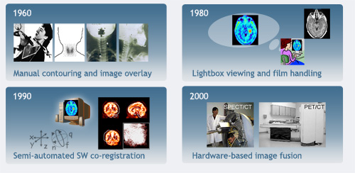
Since the 1990's, hybrid imaging by means of software and hardware image fusion alike allows the intrinsic combination of functional and anatomical image information. The introduction of hybrid imaging is predated by the invention and subsequent realization of various imaging technologies:
the discovery of X-rays by Wilhelm Conrad Roentgen in 1895, tomographic imaging with radionuclides in 1963, the introduction of the first X-ray computed tomography (CT) system in the early 1970’s, the first human tomographic images with positron-emitting isotopes were presented in 1972 and the appearance of clinical magnetic resonance (MR) in the 1980’s, to name a few milestones.
Various ways of imaging have become available over the past century that made patient observation, disease diagnosis and therapy follow-up feasible, and above all non-invasive. We know that disease originates from physical distress as well as from changes on the molecular and physiological level. In most cases of deadly diseases early diagnosis is key and, therefore, imaging the anatomy of a patient may not suffice in making a correct and timely diagnosis. Therefore, medical doctors typically employ a combination of imaging techniques during the course of diagnosis and subsequent treatment to monitor their patients. In other words, both functional and anatomical information are essential in state-of-the-art patient management.
 The advantages of integrated, anato-metabolic, or hybrid imaging are manifold. First, a single examination would provide comprehensive information on the state of a disease. Here, functional, and thus, less anatomically accurate information would be gathered and displayed in a widely appreciated anatomical context. Second, patients would be invited for only one, instead of two or multiple exams. Third, while engineering costs for combined imaging devices may initially be high, customers would benefit from purchasing a single rather than two independent devices. Fourth, the combination of complementary imaging modalities can yield synergy effects for the acquisition and processing of image data. Fifth, integrated reports on the patient can be generated based on joint expert reading from radiology and nuclear medicine physicians.
 Following its introduction into the clinic in the late 1990’s combined PET/CT imaging is now playing an increasingly important role in the diagnosis and staging of human disease.